

■ Understanding thrombosis in a graph
Under normal circumstances, there are coagulation system and anticoagulation system fibrinolysis system in the blood, and a dynamic balance is maintained between the two, so as to ensure that the blood circulates in the cardiovascular system in a liquid constant flow state.
Once the balance is broken, two clinical situation should be considered: first, whether the patient has a tendency to bleed; second, whether the patient has the risk of thrombosis. If there is bleeding tendency, plasma, platelets, cryoprecipitate may need to be supplied clinically; if there is a risk of thrombosis, heparin, anticoagulant may need to be supplied.
What is the basic physiological process of pathological thrombosis?
Due to the continuous activation of coagulation factors in the blood, a small amount of thrombin is generated to form a small amount of fibrin, which is deposited on the intima of the blood vessel, and then dissolved by the activated fibrinolytic system.
At the same time, the activated coagulation factors are also continuously phagocytosed and cleared by the mononuclear macrophage system. However, under pathological conditions, the dynamic balance between coagulation and anticoagulation is disrupted, the activity of the coagulation system is dominant, and blood coagulates in the cardiovascular system will form thrombus.
Among them, thrombotic diseases is the most concerned blood coagulation system diseases, which have the characteristics of high fatality rate, high morbidity rate and high misdiagnosis rate.
■ Application of thrombelastography in the direction of thrombosis and bleeding disorders
Thrombelastography (TEG) is a whole blood detection method which detect the beginning of coagulation to the whole process of blood clot formation and fibrinolysis, and its clinical application is increasing.
Compared with traditional coagulation testing, thromboelastography can comprehensively analyze the influence of plasma components and blood cell components on coagulation, and can more truly reflect the overall picture of coagulation in the patient's body, so as to fully consider the final effect of quantity and function on blood clot formation. It is helpful for clinicians to obtain effective information in a timely manner, and to take reasonable treatment measures for the treatment of patients with thrombosis and bleeding diseases.
1. The development history of TEG
Thromboelastography (TEG) was invented by German Hartert in 1948. It was not until 1980 that it achieved good results in clinical guidance of intraoperative blood transfusion and was widely used.Now it has become one of the most important indicators for monitoring coagulation function in the perioperative period.
In 1995, thromboelastography (TEG) began to be used in cardiac surgery. It has now become the main coagulation monitoring program during cardiopulmonary bypass and has been adopted by more than 40 countries. In 2000, it was used to guide blood coagulation-related drugs and intraoperative blood component transfusions.
Four years later, platelet mapping for monitoring the efficacy of antiplatelet drugs has brought rapid and accurate monitoring of platelet aggregation function to the clinic. In 2006, clinical laboratories adopted thromboelastography (TEG) as a screening and auxiliary diagnostic method for coagulation monitoring to provide an objective basis for clinical selection of blood products.
2. The principle and evaluation of thromboelastography (TEG) and the clinical significance of detection parameters
The principle of thromboelastography (TEG) detection is that when the test cup containing the blood sample rotates at an angle of 4°45´ and a speed of 1 cycle every 9 seconds, the coagulation strength is gradually formed with the twisting wire movement until it is almost fixed at the Moment of maximum clot stability. Once the thrombus is formed, the metal probe placed in the blood sample detection cup is subjected to the shear stress in the coagulation-thrombosis process of the sample, and then rotates left and right, and the metal needle generates current by cutting magnetic lines of force during the rotating process. After processing, the characteristic map of the movement trajectory during the period from the formation of the clot to the maximum stability of the thrombus will be formed . By comparing the shape of the trace and the results of various parameters with the normal situation, the coagulation state of the specimen can be obtained.
Thromboelastography (TEG) detection parameters include coagulation reaction time, blood clot formation time, and maximum fibrin clot strength.
Thromboelastography (TEG) as a point-of-care test is widely used in various clinical fields, such as cardiac surgery, liver transplantation, etc. Compared with traditional coagulation methods, it has the following advantages:
① The requirements for specimens are relatively low, and there is no need to process blood samples, such as whole blood, plasma, platelet-rich plasma, etc.;
②The operation is simpler and easier, no need to consider the process of specimen centrifugation, and the requirements for the operator's professional and technical level are also low;
③ The results can be qualitative and quantitative, and the computer automatically generates a variety of results, with good repeatability;
④Monitor the whole continuous process of coagulation and fibrinolysis, which is more comprehensive;
⑤The whole process only takes 20-30 minutes, which is faster;
⑥ Each detection parameter has clear clinical significance and preliminary diagnostic function, which can prompt the treatment plan for doctors.
3. Renderbio's high-throughput automatic thromboelastography opens a new chapter in the history of accurate detection
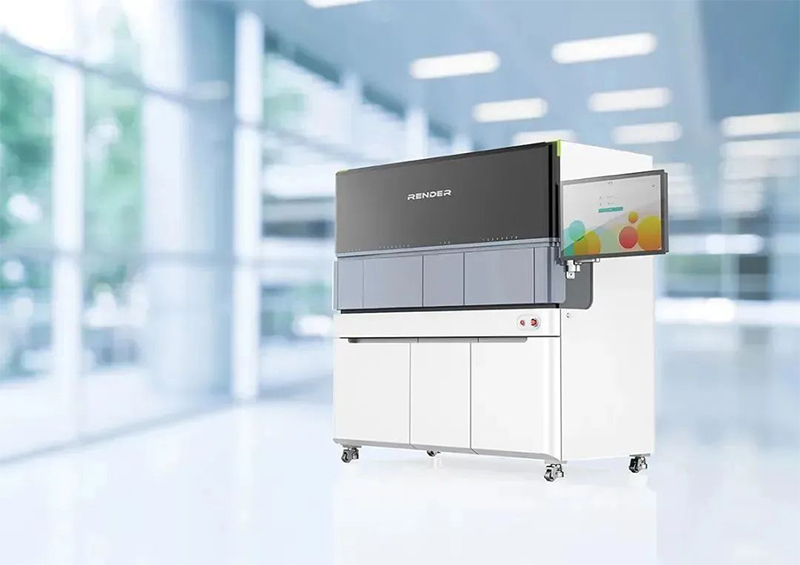
The IHTEGE12 high-throughput automatic thromboelastography instrument has 12 independent detection channels for completely parallel detection, and can process more than 200 samples per day, mainly for large hospitals with large sample volumes. Its features:
1) Adopt the most classic hanging wire method and the principle of electromagnetic cutting;
2) The whole process is automated, one-click direct access from sample to result, without manual intervention;
3) Microfluidic patented technology, pre-packaged single reagent card, ready to use;
4) No Liquid system, using disposable consumables to avoid cross-contamination and reagent waste;
5) It can be connected to the automatic assembly line of the laboratory;
6) The original tube is closed for sample injection, and the whole process is automated to ensure biological safety.